The aim of this review is to review the main pathophysiological mechanisms of bioprosthesis’ degeneration, the current interventions, either conventional or transcatheter therapies, and the future perspectives of bioengineering tissues in the degeneration of the bioprosthesis. Bioprosthesis are primarily used in valve replacements, both because they eliminate the need for oral anticoagulation and because of the specific profile of patients with valvular heart disease (elderly with higher risks of bleeding due to oral anticoagulation with warfarin, mandatory on mechanical heart valve prosthesis). However, bioprosthesis have limited durability and degeneration occurs due to the following factors: the bioprosthetic's heterologous tissue shows throughout time deposition of crystals of calcium phosphate, favored by the remnants of dead cells and fibrous structures of the tissue, resulting in dystrophic calcification; mechanical factors, since the assembly and design of the biorpothesis favors greater shear stress on the heterologous pericardial leaflets, compared to the native valve; and also to factors related to the patient, such as hypertension, left ventricular hypertrophy and patient-prosthesis mismatch (which enhances shear stress), and age (under 60 years of age), rheumatic diseases, excessive calcium excretion and up-regulation of angiotensin-coverting enzyme activity (which enhances formation of crystals of calcium phosphate). In this context, conventional reoperation for degenerated bioprosthesis is indicated; still, reoperation, especially in older patients with comorbidities, can add significant surgical risk. Transcatheter therapy (valve-in-valve and sequential valve-in-valve) emerges as recent, expanding and a viable alternative, in which a transcatheter valve is implanted within a degenerated bioprosthesis. Additionally, biological tissue engineering may enable longer-lasting bioprosthesis in the future. Tissue derived from autologous cells or pluripotent cells with decellularized xenogenic tissues may represent greater durability for bioprostheses, but require further researches and does not solve the main problem: the inexorable process of bioprothesis’ degeneration.
| Published in | Cardiology and Cardiovascular Research (Volume 9, Issue 4) |
| DOI | 10.11648/j.ccr.20250904.16 |
| Page(s) | 153-158 |
| Creative Commons |
This is an Open Access article, distributed under the terms of the Creative Commons Attribution 4.0 International License (http://creativecommons.org/licenses/by/4.0/), which permits unrestricted use, distribution and reproduction in any medium or format, provided the original work is properly cited. |
| Copyright |
Copyright © The Author(s), 2025. Published by Science Publishing Group |
Heterograft Bioprothesis, Xenograft Bioprothesis, Phisiopatholgy, Transcatheter Valve Implantation, Heart Valves
| [1] | Otto CM, Nishimura RA, Bonow RO, Carabello BA, Erwin JP 3rd, Gentile F, Jneid H, Krieger EV, Mack M, McLeod C, O’Gara PT, Rigolin VH, Sundt TM 3rd, Thompson A, Toly C. 2020 ACC/AHA Guideline for the Management of Patients With Valvular Heart Disease: Executive Summary: A Report of the American College of Cardiology/American Heart Association Joint Committee on Clinical Practice Guidelines. Circulation. 2021 Feb 2;143(5): e35-e71. Epub 2020 Dec 17. Erratum in: Circulation. 2021 Feb 2; 143(5): e228. Erratum in: Circulation. 2021 Mar 9; 143(10): e784. |
| [2] | Alec Vahanian and others, 2021 ESC/EACTS Guidelines for the management of valvular heart disease: Developed by the Task Force for the management of valvular heart disease of the European Society of Cardiology (ESC) and the European Association for Cardio-Thoracic Surgery (EACTS), European Heart Journal, Volume 43, Issue 7, 14 February 2022, Pages 561–632. |
| [3] | Banbury MK, Cosgrove DM 3rd, White JA, Blackstone EH, Frater RW, Okies JE. Age and valve size effect on the long-term durability of the Carpentier-Edwards aortic pericardial bioprosthesis. Ann Thorac Surg. 2001 Sep; 72(3): 753-7. |
| [4] | Isaacs AJ, Shuhaiber J, Salemi A, Isom OW, Sedrakyan A. National trends in utilization and in-hospital outcomes of mechanical versus bioprosthetic aortic valve replacements. J Thorac Cardiovasc Surg. 2015 May; 149(5): 1262-9. e3. |
| [5] | Huygens SA, Mokhles MM, Hanif M, Bekkers JA, Bogers AJ, Rutten-van Mölken MP, Takkenberg JJ. Contemporary outcomes after surgical aortic valve replacement with bioprostheses and allografts: a systematic review and meta-analysis. Eur J Cardiothorac Surg. 2016 Oct; 50(4): 605-616. Epub 2016 Mar 29. PMID: 27026750. |
| [6] | Forcillo J, Pellerin M, Perrault LP, Cartier R, Bouchard D, Demers P, Carrier M. Carpentier-Edwards pericardial valve in the aortic position: 25-years experience. Ann Thorac Surg. 2013 Aug; 96(2): 486-93. |
| [7] | Johnston DR, Soltesz EG, Vakil N, Rajeswaran J, Roselli EE, Sabik JF 3rd, Smedira NG, Svensson LG, Lytle BW, Blackstone EH. Long-term durability of bioprosthetic aortic valves: implications from 12,569 implants. Ann Thorac Surg. 2015 Apr; 99(4): 1239-47. |
| [8] | Kostyunin AE, Yuzhalin AE, Rezvova MA, Ovcharenko EA, Glushkova TV, Kutikhin AG. Degeneration of Bioprosthetic Heart Valves: Update 2020. J Am Heart Assoc. 2020 Oct 20; 9(19): e018506. |
| [9] | Kostyunin AE, Yuzhalin AE, Ovcharenko EA, Kutikhin AG. Development of calcific aortic valve disease: Do we know enough for new clinical trials? J Mol Cell Cardiol. 2019 Jul; 132: 189-209. |
| [10] | Simionescu DT. Prevention of calcification in bioprosthetic heart valves: challenges and perspectives. Expert Opin Biol Ther. 2004 Dec; 4(12): 1971-85. |
| [11] | Dalgliesh AJ, Parvizi M, Noble C, Griffiths LG. Effect of cyclic deformation on xenogeneic heart valve biomaterials. PloS One. 2019 Jun 13; 14(6): e0214656. |
| [12] | Tam H, Zhang W, Feaver KR, Parchment N, Sacks MS, Vyavahare N. A novel crosslinking method for improved tear resistance and biocompatibility of tissue based biomaterials. Biomaterials. 2015 Oct; 66: 83-91. |
| [13] |
Vyavahare N, Ogle M, Schoen FJ, Zand R, Gloeckner DC, Sacks M, Levy RJ. Mechanisms of bioprosthetic heart valve failure: fatigue causes collagen denaturation and glycosaminoglycan loss. J Biomed Mater Res. 1999 Jul; 46(1): 44-50.
https://doi.org/10.1002/(sici)1097-4636(199907)46:1<44::aid-jbm5>3.0.co;2-d |
| [14] | Ghaisas NK, Foley JB, O’Briain DS, Crean P, Kelleher D, Walsh M. Adhesion molecules in nonrheumatic aortic valve disease: endothelial expression, serum levels and effects of valve replacement. J Am Coll Cardiol. 2000 Dec; 36(7): 2257-62. |
| [15] | Singhal P, Luk A, Butany J. Bioprosthetic Heart Valves: Impact of Implantation on Biomaterials. ISRN Biomaterials 2013; 1-14. |
| [16] | Sturla F, Ronzoni M, Vitali M, Dimasi A, Vismara R, Preston-Maher G, Burriesci G, Votta E, Redaelli A. Impact of different aortic valve calcification patterns on the outcome of transcatheter aortic valve implantation: A finite element study. J Biomech. 2016 Aug 16; 49(12): 2520-30. |
| [17] | Abbasi M, Azadani AN. Leaflet stress and strain distributions following incomplete transcatheter aortic valve expansion. J Biomech. 2015 Oct 15; 48(13): 3663-71. |
| [18] | Bourget JM, Zegdi R, Lin J, Wawryko P, Merhi Y, Convelbo C, Mao J, Fu Y, Xu T, Merkel NO, Wang L, Germain L, Zhang Z, Guidoin R. Correlation between structural changes and acute thrombogenicity in transcatheter pericardium valves after crimping and balloon deployment. Morphologie. 2017 Mar; 101(332): 19-32. |
| [19] | Martin C, Sun W. Comparison of transcatheter aortic valve and surgical bioprosthetic valve durability: A fatigue simulation study. J Biomech. 2015 Sep 18; 48(12): 3026-34. |
| [20] | Søndergaard L, Ihlemann N, Capodanno D, Jørgensen TH, Nissen H, Kjeldsen BJ, Chang Y, Steinbrüchel DA, Olsen PS, Petronio AS, Thyregod HGH. Durability of Transcatheter and Surgical Bioprosthetic Aortic Valves in Patients at Lower Surgical Risk. J Am Coll Cardiol. 2019 Feb 12; 73(5): 546-553. |
| [21] | Leon MB, Smith CR, Mack M, Miller DC, Moses JW, Svensson LG, Tuzcu EM, Webb JG, Fontana GP, Makkar RR, Brown DL, Block PC, Guyton RA, Pichard AD, Bavaria JE, Herrmann HC, Douglas PS, Petersen JL, Akin JJ, Anderson WN, Wang D, Pocock S; PARTNER Trial Investigators. Transcatheter aortic-valve implantation for aortic stenosis in patients who cannot undergo surgery. N Engl J Med. 2010 Oct 21; 363(17): 1597-607. |
| [22] | Cribier A, Eltchaninoff H, Bash A, Borenstein N, Tron C, Bauer F et al. Percutaneous transcatheter implantation of an aortic valve prosthesis for calcific aortic stenosis: first human case description. Circulation 2002; 106(24): 3006-3008. |
| [23] | Wenaweser P, Buellesfeld L, Gerckens U, Grube E. Percutaneous aortic valve replacement for severe aortic regurgitation in degenerated bioprosthesis: the first valve in valve procedure using the Corevalve Revalving system. Catheter Cardiovasc Interv. 2007 Nov 1; 70(5): 760-4. |
| [24] | Leung Wai Sang S, Giri J, Vallabhajosyula P. Transfemoral transcatheter valve-in-valve-in-valve replacement. J Thorac Cardiovasc Surg. 2016 Aug; 152(2): 622-3. |
| [25] | Cardoso CC, Gaia DF, Palma JH, Oliveira Jr JL. The End of Repeated Reoperations for Degenerated Bioprosthesis? Hydrodynamic In-Vitro Analysis for Sequential Valve-in-Valve Feasibility. J Clin Exp Cardiolog. 2024 Sep; 15: 912. |
| [26] | Shinoka T. Tissue engineered heart valves: autologous cell seeding on biodegradable polymer scaffold. Artif Organs. 2002 May; 26(5): 402-6. |
| [27] | V. K. Bajpai, S. T. Andreadis. Stem cell sources for vascular tissue engineering and regeneration. T issue Eng. Part B Rev., 18 (5) (2012), pp. 405-425. |
| [28] | Filová E, Straka F, Miřejovský T, Mašín J, Bačáková L. Tissue-engineered heart valves. Physiol Res. 2009; 58 Suppl 2: S141-S158. |
APA Style
Cardoso, C. C., Peres, A. L. B. (2025). Degeneration of Biological Heart Valve Prosthesis: Review of Pathophysiological Mechanisms, Current Interventions and Future Perspectives. Cardiology and Cardiovascular Research, 9(4), 153-158. https://doi.org/10.11648/j.ccr.20250904.16
ACS Style
Cardoso, C. C.; Peres, A. L. B. Degeneration of Biological Heart Valve Prosthesis: Review of Pathophysiological Mechanisms, Current Interventions and Future Perspectives. Cardiol. Cardiovasc. Res. 2025, 9(4), 153-158. doi: 10.11648/j.ccr.20250904.16
AMA Style
Cardoso CC, Peres ALB. Degeneration of Biological Heart Valve Prosthesis: Review of Pathophysiological Mechanisms, Current Interventions and Future Perspectives. Cardiol Cardiovasc Res. 2025;9(4):153-158. doi: 10.11648/j.ccr.20250904.16
@article{10.11648/j.ccr.20250904.16,
  author = {Caio Cesar Cardoso and Ana Luiza Boucault Peres},
  title = {Degeneration of Biological Heart Valve Prosthesis: Review of Pathophysiological Mechanisms, Current Interventions and Future Perspectives
},
  journal = {Cardiology and Cardiovascular Research},
  volume = {9},
  number = {4},
  pages = {153-158},
  doi = {10.11648/j.ccr.20250904.16},
  url = {https://doi.org/10.11648/j.ccr.20250904.16},
  eprint = {https://article.sciencepublishinggroup.com/pdf/10.11648.j.ccr.20250904.16},
  abstract = {The aim of this review is to review the main pathophysiological mechanisms of bioprosthesis’ degeneration, the current interventions, either conventional or transcatheter therapies, and the future perspectives of bioengineering tissues in the degeneration of the bioprosthesis. Bioprosthesis are primarily used in valve replacements, both because they eliminate the need for oral anticoagulation and because of the specific profile of patients with valvular heart disease (elderly with higher risks of bleeding due to oral anticoagulation with warfarin, mandatory on mechanical heart valve prosthesis). However, bioprosthesis have limited durability and degeneration occurs due to the following factors: the bioprosthetic's heterologous tissue shows throughout time deposition of crystals of calcium phosphate, favored by the remnants of dead cells and fibrous structures of the tissue, resulting in dystrophic calcification; mechanical factors, since the assembly and design of the biorpothesis favors greater shear stress on the heterologous pericardial leaflets, compared to the native valve; and also to factors related to the patient, such as hypertension, left ventricular hypertrophy and patient-prosthesis mismatch (which enhances shear stress), and age (under 60 years of age), rheumatic diseases, excessive calcium excretion and up-regulation of angiotensin-coverting enzyme activity (which enhances formation of crystals of calcium phosphate). In this context, conventional reoperation for degenerated bioprosthesis is indicated; still, reoperation, especially in older patients with comorbidities, can add significant surgical risk. Transcatheter therapy (valve-in-valve and sequential valve-in-valve) emerges as recent, expanding and a viable alternative, in which a transcatheter valve is implanted within a degenerated bioprosthesis. Additionally, biological tissue engineering may enable longer-lasting bioprosthesis in the future. Tissue derived from autologous cells or pluripotent cells with decellularized xenogenic tissues may represent greater durability for bioprostheses, but require further researches and does not solve the main problem: the inexorable process of bioprothesis’ degeneration.
},
 year = {2025}
}
TY  - JOUR
T1  - Degeneration of Biological Heart Valve Prosthesis: Review of Pathophysiological Mechanisms, Current Interventions and Future Perspectives

AU  - Caio Cesar Cardoso
AU  - Ana Luiza Boucault Peres
Y1  - 2025/12/03
PY  - 2025
N1  - https://doi.org/10.11648/j.ccr.20250904.16
DO  - 10.11648/j.ccr.20250904.16
T2  - Cardiology and Cardiovascular Research
JF  - Cardiology and Cardiovascular Research
JO  - Cardiology and Cardiovascular Research
SP  - 153
EP  - 158
PB  - Science Publishing Group
SN  - 2578-8914
UR  - https://doi.org/10.11648/j.ccr.20250904.16
AB  - The aim of this review is to review the main pathophysiological mechanisms of bioprosthesis’ degeneration, the current interventions, either conventional or transcatheter therapies, and the future perspectives of bioengineering tissues in the degeneration of the bioprosthesis. Bioprosthesis are primarily used in valve replacements, both because they eliminate the need for oral anticoagulation and because of the specific profile of patients with valvular heart disease (elderly with higher risks of bleeding due to oral anticoagulation with warfarin, mandatory on mechanical heart valve prosthesis). However, bioprosthesis have limited durability and degeneration occurs due to the following factors: the bioprosthetic's heterologous tissue shows throughout time deposition of crystals of calcium phosphate, favored by the remnants of dead cells and fibrous structures of the tissue, resulting in dystrophic calcification; mechanical factors, since the assembly and design of the biorpothesis favors greater shear stress on the heterologous pericardial leaflets, compared to the native valve; and also to factors related to the patient, such as hypertension, left ventricular hypertrophy and patient-prosthesis mismatch (which enhances shear stress), and age (under 60 years of age), rheumatic diseases, excessive calcium excretion and up-regulation of angiotensin-coverting enzyme activity (which enhances formation of crystals of calcium phosphate). In this context, conventional reoperation for degenerated bioprosthesis is indicated; still, reoperation, especially in older patients with comorbidities, can add significant surgical risk. Transcatheter therapy (valve-in-valve and sequential valve-in-valve) emerges as recent, expanding and a viable alternative, in which a transcatheter valve is implanted within a degenerated bioprosthesis. Additionally, biological tissue engineering may enable longer-lasting bioprosthesis in the future. Tissue derived from autologous cells or pluripotent cells with decellularized xenogenic tissues may represent greater durability for bioprostheses, but require further researches and does not solve the main problem: the inexorable process of bioprothesis’ degeneration.

VL  - 9
IS  - 4
ER  -
Faculty of Medical Sciences of the Santa Casa of Sao Paulo, Sao Paulo, Brazil
Biography: Caio Cesar Cardoso is a physician graduated from the Faculty of Medical Sciences of Santa Casa de Sao Paulo. General Surgeon from the Irmandade of Santa Casa de Misericórdia de Sao Paulo and Cardiovascular Surgeon from the Federal University of Sao Paulo. Has professional Master's Degree in New Technologies and Healthcare from the Federal University of Sao Paulo and doctorate in Cardiology from the Federal University of Sao Paulo. Works as professor of Medicine at the School of Medical Sciences of Santa Casa de Sao Paulo, physician in charge of the Cardiac Implantable Electronic Devices Outpatient Clinic of the Irmandade of Santa Casa de Misericórdia de Sao Paulo, head of the Cardiovascular Surgery Department at the Adventist Hospital of Sao Paulo and Cardiovascular Surgeon at Stella Maris Hospital. Also is an Associate Editor of Journal of Cardiology and Cardiovascular Research.
Research Fields: cardiovascular surgery, transcatheter valves, valve-in-valve, bioprosthesis, heart valve prosthesis

Faculty of Medical Sciences of the Santa Casa of Sao Paulo, Sao Paulo, Brazil
Biography: Ana Luiza Boucault Peres is a medical student at the Faculty of Medical Sciences of Santa Casa de Sao Paulo, where she began her studies in 2024. She aims to contribute to the advancement of evidence-based medicine and the improvement of patient care through continuous learning and research involvement.
Research Fields: medicine, heart valve prosthesis

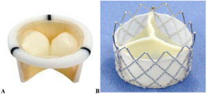
Figure 1. A. Braile conventional bioprosthesis. B. Braile Inovare transcatheter valve.
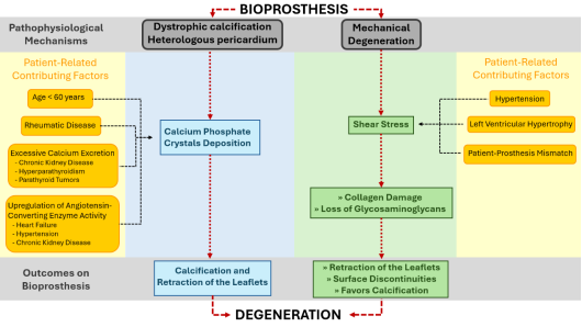
Figure 2. Diagram illustrating the pathophysiological mechanisms involved in the degeneration of bioprosthesis.
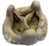
Figure 3. Bioprosthesis with intense degeneration, highlighting calcification of the cusps. Extracted from Kostyunin et al 2020.
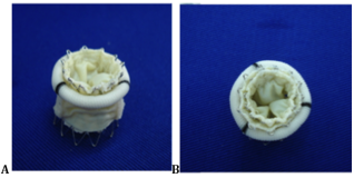
Figure 4. A. A set of a Braile conventional bioprosthesis and a Braile Inovare transcatheter valve. B. The implantation of another Braile Inovare transcatheter valve inside the set shown in A.